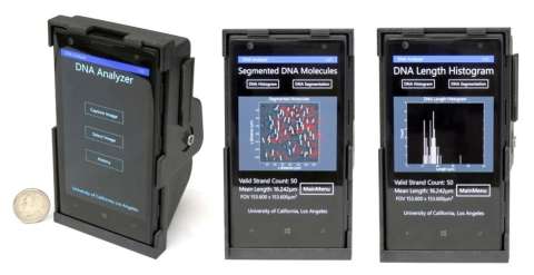
 Researchers at University of California, Los Angeles (UCLA) have developed a new technology that turns a smartphone into a DNA-scanning fluorescent microscope.
Lead researcher Aydogan Ozcan, Howard Hughes Medical Institute chancellor professor at UCLA, sat down with Bioscience Technology to talk about this advancement and its implications for resource-poor labs, and for personalized medicine.
The new optical attachment, which includes a lens, filter, mount and laser diode in a 3D-printed case, can image and size DNA molecules 50,000 times thinner than a human hair.
Scientists see the technology being used in remote laboratory settings to diagnose cancers and central nervous system disorders such as Alzheimer’s, and to detect drug resistance in infectious diseases.
Bringing techniques and testing that is normally confined to a laboratory or hospital, out into the field, or right into a patient’s home is a theme in Ozcan’s lab.
His lab, “is working on computational optical technologies that aim for microscopy, imaging, sensing and diagnostic applications, unlike traditional techniques that are using instruments that you normally find in a lab or hospital,” he said. “We’re doing it using interfaces that are simple, lightweight, compact and cost effective, that utilize consumer devices (especially mobile phones) as a platform for making these measurements in filed settings and resource-limited settings.”
Here’s how it works.
First, the desired DNA must be isolated and labeled with fluorescent tags, a procedure that can be done even in remote locations or limited resources, Ozcan, who leads the Bio- and Nano-photonics Laboratory at UCLA Electrical Engineering and Bioengineering Departments, said. To scan the DNA researchers developed a computational interface and Windows smart application running on the same smartphone. Information is then sent to a remote server in the researchers’ UCLA laboratory that measures the length of the DNA molecules. The data processing can take less than 10 seconds depending on the data connection.
Read More: How a Smartphone Camera Can Find Eye Cancer
Traditional microscopes can do the same thing – but they are bulky, expensive and often unavailable in remote locations. Not only does this technology reduce cost, and enable telemedicine and mobile health, but there’s also another angle that makes them attractive, Aydogan said, and that is connectivity.
Unlike traditional microscopes, which are used locally and in a disconnected fashion, the microscopes described here are all connected to servers through WIFI or network signals, which make them “quite powerful” in terms of labeling results as a function of space and time and see the spreading of certain conditions or identify a trend. In HIV, for example, “you can ask a question like, how is that expanding or spreading over the last year compared to the previous year, or why is it increasing in this neighborhood versus another one,” Ozcan explained.
The attachment reliably sized DNA segments of 10,000 base pairs or longer, a size range that many important genes fall in, according to the researchers. This includes a bacterial gene known for giving staphylococcus aureus and other bacteria antibiotic resistance that is about 14,000 base pairs long.
For 5,000 base-pair or shorter segments, however, there was a significant drop in accuracy due to the reduced detection signal-to-noise ratio and contrast for such short fragments. One solution to this problem is to replace the attachment’s current lens with one that has a higher numerical aperture, the researchers said.
Variety of Uses
In general, the smartphone technology being developed at Ozcan’s lab can be used to perform a number of lab functions, such as diagnosing and tracking Malaria and TB. It can also be applied to blood diseases, like sickle cell anemia, or be used to look at contamination, for example in food or milk. The team has been able to convert the mobile phone into a sensitive E-coli or giardia detector, one of the most frequently encountered pathogens, Ozcan said. It can also be used for simple tests that are normally only done at hospitals, such as total count of red or white blood cells.
In the home and in the field
Doctors in the field, “can convert a simple nurse office or a point of care office into an advanced testing infrastructure,” Ozcan said. “They can, for example, look at a Malaria infected patient, or TB infected patient and potentially decide on a drug choice based on some of the genetic testing copy number variations of certain genes that you would find in the sample taken from the patient.”
The technology also removes barriers to testing that cities or small villages might have, including the cost of shipping and sending of specimen, or lack of experts in the immediate area. “If you were to have these microscopes that are extremely cost effective, a simple nurse or a healthcare technician can prepare specimen and image them, where the images are then transferred to an expert professional pathologist that is maybe 1,000 miles away or maybe not even in the same country.”
One way this technology could be used in places like the US, is for chronic patients or the aging population. “Rather than bringing these patients out of their homes, you bring the lab to the home and do testing extremely frequently.” For example, someone with diabetes who has chronic kidney problems. If the person needed to be tested every few hours, before a meal, after a meal, it would be very valuable information for your doctor to be able to track your condition, Ozcan said. “I think that it’s a great opportunity, especially for lowering insurance costs in the US when it becomes more of a problem to manage our chronic patients and our aging population.”
Next up the researchers plan to test their device in the field to detect the presence of malaria-related drug resistance. The team also has other devices in the pipeline that they are currently testing and comparing their performance against lab instruments that are FDA approved.
The research, “Field-Portable Smartphone Microscopy Platform for Wide-field Imaging and Sizing of Single DNA Molecules,” was presented at the Optical Society’s Conference on Laser and Electro Optics (CLEO) 2015.


