Articles
LISTED UNDER:
 Optogenetics is revolutionizing the field of behavioral neuroscience by enabling researchers to control the activity of individual neurons and measure the effects of those manipulations. Hundreds of labs are using the technique to explore the neurobiology of phenomena such as decision-making and neurodegenerative diseases, often with remarkable results.
Fluorescent imaging is a widely used technique to gain insight into biological functions. But problems can arise when external illumination is required, as in optogenetics: The same light that stimulates what's under optogenetic control can interfere with the fluorescent sensor. For instance, the same blue light that excites the FRET-based indicator for calcium activates a commonly used photo-sensitive receptor used in optogenetics.
Light can be generated instead by chemiluminescence– where the emission is produced by a chemical reaction– but existing chemiluminescent probes are too weak for use in optogenetics studies. A means to create brighter chemiluminescence would help to advance these and other applications.
To investigate the input-output relation in living cells, compatible use of optogenetics and fluorescent indicators such as calcium ion (Ca2+) are very useful combinations. However, because fluorescent indicators require excitation light sources, such excitation light gives perturbations to the cell function as photo-stimulation light.
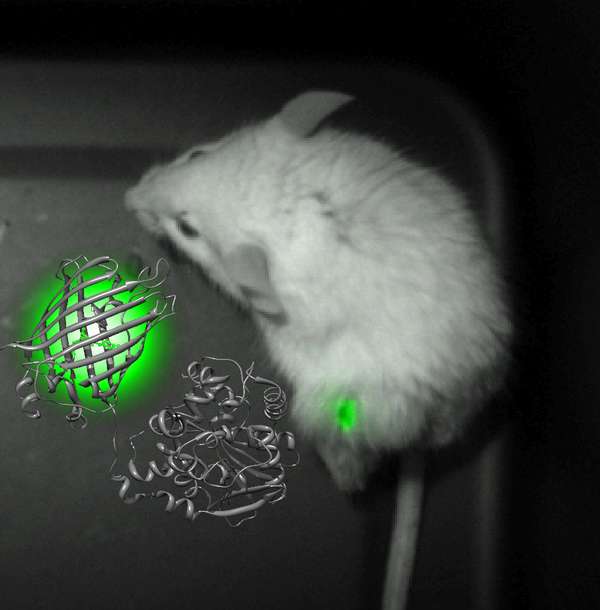
To overcome this problem, a lab at Osaka University, Japan, engineered a probe that fuses a luminescent protein from the sea pansy Renilla reniformis with a fluorescent protein we previously designed. The new molecular entity – dubbed Nano-lantern – is considered the world's brightest luminescent protein, with temporal and spatial resolution equivalent to those of fluorescence.
Dispensing with the need for light illumination, which is essential for fluorescence imaging, allowed us to analyze dynamics in environments where the use of fluorescent indicators is not feasible.
Once the new protein was designed, which is 10 times brighter than the original template, it was put through its paces. First, we used it to image intracellular structures in living cells, demonstrating spatial resolution and brightness on par with those of fluorescence.
Collaborating with Dr. Yuriko Higuchi at Kyoto University, the research team from Osaka University visualized cancer tissue inside a freely moving mouse. Here, the Nano-lantern offered a number of advantages over conventional probes, including increased sensitivity and shorter exposure times. It even enabled video-rate imaging of tumors 17 days after implantation.
Next, the group modified the Nano-lantern into a calcium sensor and co-expressed it with a light-sensitive photoreceptor in rat neurons. Fluorescent Ca2+ indicators by themselves would be triggered by light, making it impossible to perform optogenetics. This co-expression system allowed the researchers to follow excitation of the photoreceptors by measuring the Ca2+ increase as reported by the Nano-lantern Ca2+ indicator.
The group was also able to visualize ATP production in plant chloroplasts. They converted the Nano-lantern into an ATP sensor, and expressed it in a live leaf. The red autofluorescence signal of the chloroplasts did not interfere with the Nano-lantern, allowing observation of the increase in ATP levels after light irradiation – as well as the first visualization of the qE quenching-mediated down regulation of ATP synthesis in live plant cells.
To visualize Nano-lantern signals, the researchers used Photometrics' Evolve 512 EMCCD camera because they knew that the Photometrics' cameras worked very well, having used the Cascade II EMCCD before trying the Evolve.
The Evolve's high quantum efficiency allowed the research team to easily detect the low chemiluminescent signals of RLuc for both still images and video-rate imaging without the risk of phototoxicity. The camera's superior cooling (-85°C) increased the signal-to-noise ratio for the mouse tumor imaging, while its wide dynamic range enabled acquisition of both chemiluminescent and bright-field images using the same camera setting.
The Evolve 512 EMCCD also offers a feature that allowed the team to perform "cleaner" optogenetic experiments. Because the light used to stimulate optogenetic processes is so strong, it can increase the background noise level. To overcome this issue, the team used the dead-time of the Evolve 512 camera to conduct optogenetic light stimulation.
One of the unique advantages of Evolve 512 is that it can erase charges on the CCD during the dead-time. This function contributed to reduced background noise for compatible use of optogenetic and chemiluminescent imaging during the research.
Now, all biological phenomena that are regulated by light, including photosynthesis and photoreception in retina, can be viewed using Nano-lantern, giving researchers a more dynamic and quantitative analysis of living cells.

