Multi-Parametric Electrophysiological Imaging of the Mammalian Heart in vivo
LISTED UNDER:
 Cardiac arrhythmia is one of the most common diseases encountered in clinical cardiology. High-speed electrophysiological imaging using fluorescent probes has yielded tremendous insights into the basic mechanisms of arrhythmias and the effects of anti-arrhythmic drugs. However, optical mapping, as it is known to the cardiac research community, has remained relegated to the isolated (i.e. explanted) heart. Although useful, the traditional Langendorff-perfused isolated heart preparation is unphysiological in many respects. For instance, in this system, the heart is explanted from the animal, disconnecting neuro-humeral feedback, and is perfused with saline, a condition lethal to the live animal. Hence, in vivo optical mapping must be developed.
Two key parameters of interest are the cardiac action potential (AP) and intracellular calcium transient (CaT), which requires the use of red-shifted dyes to image in vivo. Peter Lee, PhD, and a collaborator, Dr. Christopher Woods, MD PhD, cardiologist in the Division of Cardiovascular Medicine at Stanford University, demonstrated the first ever multi-parametric imaging of a mammalian heart in vivo through whole blood.
“The purpose of this in vivo study was to transition to imaging electrophysiological parameters with the heart still inside the animal through 100 percent blood,” said Lee. “We showed you can measure these two parameters that people care about in a live animal and you can do it quite simply with a single camera.”
Challenge
The Langendorff-perfused mammalian heart preparation has been used to study heart function for more than a century. In combination with optical mapping, this technique has given researchers insight into arrhythmia biology. However, the unphysiological nature of the heart preparation makes it difficult for researchers to apply this data in a clinically relevant setting.
“It is the critical neuro-humeral and hormonal influence present in patients that we believe leads to important diseases like heart failure and arrhythmia, and we simply could not study this well in the explanted heart. But, what Peter has designed here has really been transformative. For the first time, we will be able to understand these disease processes where they happen over time," notes Dr. Woods.
Traditional methods of imaging the heart in vivo, such as magnetic resonance imaging (MRI) or ultrasound, provide relatively slow, low-resolution images that lack the ability to capture electrophysiological data. Thus far, only one in vivo optical mapping study has been reported. However, the research team was limited to studying a single parameter (voltage) using a low-resolution imaging system and complex illumination system based on lasers and acousto-optic deflectors. No further progress has been reported since that 1998 study.
Prior to Lee’s research, multi-parametric imaging required the use of two cameras, making it costly and technically difficult for researchers to perform. Traditional light sources for such multi-color imaging require mechanical shuttering devices or filter wheels to quickly switch between excitation wavelengths, raising set-up complexity and component costs.
Lee and his colleagues developed a simple and scalable single-camera-imaging and LED-illumination system that reduces the economic and technical hurdles associated with in vivo imaging. They also used a red-shifted calcium dye (blood does not absorb strongly in red wavelengths) and a new near-infrared (NIR) voltage-sensitive dye, both ideal for whole blood-perfused tissue.
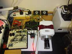
As part of his single camera, multi-parametric imaging design, Lee relied on Photometrics’ Evolve 128 EMCCD camera. His system was also comprised of off-the-shelf optical filters and lenses and powerful LED chips which enabled the change from, according to Lee, a “single light source—multi-camera approach” to a “multi light source—single camera approach.”
“For imaging particular fluorescent dyes in cardiac tissue, the camera has to have certain properties, including high speed and a large well depth. The cameras from Photometrics fit the bill quite nicely,” said Lee.
With a 10-MHz readout, the Evolve 128 was designed for high-speed image visualization. It also has the lowest read noise available for EMCCD cameras. When dealing with commonly desired cardiac physiological parameters, achieving very high speeds (as can be acquired with the Evolve 128) is not as much an issue as with other set-ups. Therefore, Lee was able to manipulate the speed by sharing the frames between samples, allowing him to conduct his research with only a single camera.
The fluorescence emission is then passed through a multi-band emission filter and camera lens. During any frame exposure period of the camera, the tissue is illuminated with only one of the excitation sources. Because of the established lack of crosstalk between the dyes, the emitted fluorescence at any time represents only one of the parameters. The multi-band filter was essential in allowing Lee to image multiple parameters along the same optical path.
Due to the high camera frame rate of ~1000 fps at 64x64 pixel resolution and smooth signal dynamics, with interpolation, one can measure all the parameters in a straightforward fashion. A high-speed microcontroller coordinates the fast-response-time LEDs with the frame exposure signal from the camera. A standard desktop computer is used to support the camera system and to communicate with the microcontroller-based controller board.
Results
Lee and Woods hope that their proof-of-concept imaging system will open the doors for clinically-relevant arrhythmia research. By adapting the system to minimally-invasive and clinically-compatible trans-catheter endoscopic methods for large animals and patients, such in vivo multi-parametric imaging will enable unprecedented high spatio-temporal electrophysiological studies of the heart in live animals, thus advancing arrhythmia treatment.


