SEM for 3-D Volume Imaging of Cells and Tissues
LISTED UNDER:
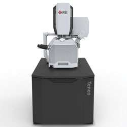
 The Teneo VS scanning electron microscope (SEM) from FEI offers a VolumeScope capability for life science applications. The Teneo platform tightly integrates FEI’s latest-generation SEM with VolumeScope, an in-chamber microtome and proprietary analytical software to provide fully-automated, large-volume reconstructions with dramatically improved z-axis resolution. VolumeScope uses serial block face imaging (SBFI) to acquire a 3-D volume from a block of tissue or cells. While SEMs can provide nanometer-scale lateral resolution in images of each slice, the axial resolution of the reconstructed model is normally limited by the physical thickness of the slices, typically 25 micrometers or more. The Teneo VS uses FEI’s ThruSight multi-energy deconvolution to resolve features at different depths within each slice, thus improving axial resolution. The VolumeScope in-chamber ultra-microtome is fully-integrated with the Teneo VS operating and imaging software. Switching between volume imaging and normal SEM operation is fast and easy. MAPS software uses tiling and stitching to acquire high-resolution composite images that are much larger than a single field of view. MAPS also permits correlation with light microscope images for easy targeting of specific areas of interests. Reconstruction, visualization, analysis and presentation are handled by Amira software, which imports the image data directly from the microscope. The system also offers a low-vacuum water vapor option with dedicated detection that can reduce charging and provide further improvements in contrast and resolution on challenging samples.
FEI, http://www.fei.com/teneo-for-life-sciences

