Simultaneously Tracking Blood Flow, Oxygenation Can Revolutionize Neural Imaging
Quantifying brain activity through optical imaging has the potential to improve the way the biomedical community treats neurological disorders and brain injuries. To accurately visualize and treat patients who have suffered a stroke, epileptic attack or traumatic brain injury, neuroscientists require precise imaging and measurements of brain activity.
Standard brain imaging techniques, such as computed tomography (CT) or magnetic resonance imaging (MRI), are primarily intended to distinguish the anatomy of the brain and detect physical abnormalities such as tumors, infarctions, scar tissue and hemorrhages, which help in diagnosing various conditions including dementia and epilepsy. These imaging modalities, however, are limited in their ability to monitor physiological events in real time, including blood flow, oxygenation, ion transport and electrical signaling. Physicians would greatly benefit from an imaging method that allows real-time tracking of brain activity to validate the effectiveness of new treatments.
One area of interest for understanding abnormal brain activity is tracking blood flow and oxygenation changes. However, measuring both neural activities requires two imaging techniques with conflicting light source properties and camera requirements. Blood oxygenation can be measured through a technique called Intrinsic Signal Optical Imaging, which involves measuring the absorption of different wavelengths of non-coherent light to determine the concentration of oxygenated versus deoxygenated hemoglobin. Conversely, blood flow is measured through a technique called Laser Speckle Contrast Imaging, which captures the random speckle pattern produced by light scattered from a coherent laser as it interacts with moving blood cells. Studying the combination of blood flow and oxygenation together is vital to a better understanding of the underlying dynamics of epilepsy and stroke. Until now, researchers have been faced with the challenge of measuring both parameters simultaneously on a single system.
.jpg) Finding the light
Finding the light
Dr. Ofer Levi, Ph.D., assistant professor at the University of Toronto’s Institute of Biomaterials and Biomedical Engineering, and his team set out to prove that with the right technology and skills, accurate, real-time, dual-modality brain blood flow and oxygenation mapping using a single system is possible.
The Levi lab uses laboratory rats to monitor the brain’s response to ischemia (a lack of blood flow to the brain). To observe these effects, the team designed a system that could accurately measure both oxygenation and blood flow in real-time.
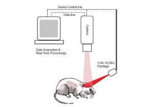
This breakthrough was achieved by working with vertical-cavity surface-emitting lasers (VCSELs) at the near infrared wavelength region (above 680 nanometers) where light penetrates deeply in the tissue. Using VCSELs as their illumination light source and modulating the current, they were able to rapidly switch between highly coherent light required for measuring blood flow and low coherent light required for measuring oxygenation. With this technique, Levi now had the ability to simultaneously measure both parameters using this single light source in real time.
Creating a custom brain mapping system
Once they identified their light source, Levi teamed up with scientific camera provider, QImaging, to create a custom optical brain imaging system. Because of Levi’s unique imaging requirements, he consulted with the QImaging team to find a camera solution that would address the different imaging requirements of these two distinct techniques.
He chose to work with QImaging for three reasons: their technical expertise, knowledge of the application and their ability to translate the luminescence and optical imaging constraints to camera requirements. Working together, QImaging and Levi were able to identify a single camera solution that would provide the required dynamic range, speed and integration with their custom light source to satisfy both imaging modalities.
Scientists often underestimate the importance of establishing a great connection with their imaging provider. Levi proved the importance of finding a team that offers the technical and application expertise required to find the right camera solution for your imaging technique, as they both deserve equal attention.
The experts at QImaging recommended Levi use the Rolera EM-C2 camera, which provides the dynamic range, high frame rates and low-read noise required to accurately measure both brain dynamics with sufficient temporal and spatial resolution. These features, combined with the precise synchronization between the camera and the VCSELs, enabled by high-speed hardware triggering from the EM-C2, allowed rapid switching between the two imaging techniques for simultaneous imaging of blood flow and oxygenation at 100 frames per second. With this camera and light source configuration, Levi’s team was able to generate in vivo blood flow and oxygenation maps of a rodent brain in response to induced ischemia.
Real-time, detailed imaging
With this novel dual-modality imaging technique, Levi’s lab was able to expand their research focus to extending the depth of field for in vivo brain imaging and studying the dynamics of blood-brain barrier (BBB) breaching due to the effects of brain disease or a drug.
Next, Levi plans to use QImaging’s new optiMOS Scientific CMOS (sCMOS) camera, which he hopes will enable faster frame rates for better temporal resolution and greater accuracy in blood flow and oxygenation maps.
Using current brain imaging systems, medical experts require six to 12 weeks to determine whether stroke or epilepsy treatments are taking effect. Looking to the future, Levi aims to translate his proof of concept system to the clinic to dramatically reduce that response time to days. Such rapid feedback would lead to more targeted therapies and allow physicians to alter treatment instantaneously.
References
- I. Sigal, R. Gad, A. M. Caravaca-Aguirre, Y. Atchia, D. B. Conkey, R. Piestun, and O. Levi, "Laser speckle contrast imaging with extended depth of field for in-vivo tissue imaging," Biomed. Opt. Express 5, 123-135 (2014).
- S. Dufour, Y. Atchia, R. Gad, D. Ringuette, I. Sigal, and O. Levi, "Evaluation of laser speckle contrast imaging as an intrinsic method to monitor blood brain barrier integrity," Biomed. Opt. Express 4, 1856-1875 (2013).
- O. Levi, "Multimodal Optical Neural Imaging using VCSELs," in Imaging and Applied Optics (Optical Society of America, Arlington, Virginia, 2013), p. ITh1D.1.
- O. Levi, H. levy, D. Ringuette, “Rapid monitoring of cerebral ischemia dynamics using laser-based optical imaging of blood oxygenation and flow,” Biomedical Optics Express, 4, 777-791 (2012)


